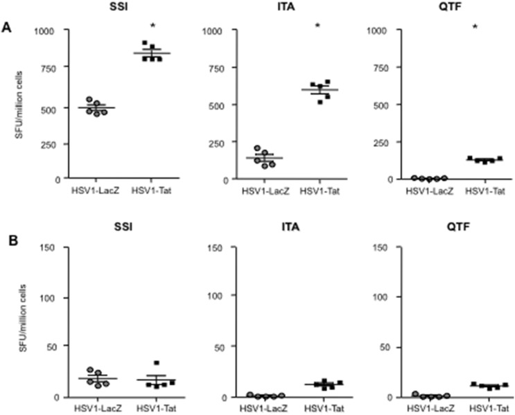
Figure 6. Analysis of HSV1-specific T-cell responses in individual C57BL/6 mice.
Mice were inoculated intravaginally with 103 pfu/mouse of live attenuated HSV1-Tat or HSV1-LacZ. Seven days after infection, splenocytes were isolated from 5 mice/group and tested by ELISpot assay for IFN-γ (A) or IL-4 (B) cytokine production upon stimulation with SSI, ITA and QTF HSV-derived peptides. Results are expressed as number of spot-forming units (SFU)/million cells per mouse. Values at least 2-fold higher than the mean number of spots in the control wells (untreated cells), i.e., ≥50 SFU/million cells, were considered positive. The results of one representative experiment (out of three) are shown. The two-tailed Mann-Whitney test was used for statistical analysis, *p<0.05.

