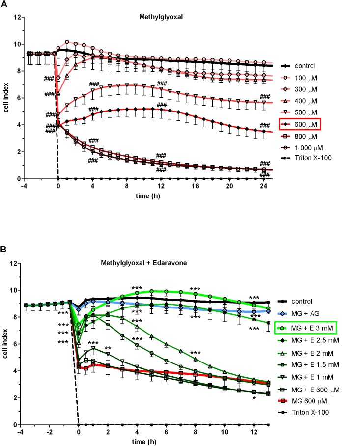
Figure 1. Effect of methylglyoxal and edaravone on cell viability.
Effect of methylglyoxal (100–1000 µM) on human hCMEC/D3 endothelial cells measured by real-time cell electronic sensing method (A). Effect of co-treament with 600 µM methylglyoxal and different concentrations of edaravone (MG + E; 600–3000 µM) or aminoguanidine (MG + AG; 2 mM) (B). Cell index is expressed as an arbitrary unit and calculated from impedance measurements between cells and sensors. Data are presented as means ± SD, n = 10. Triton X-100 was used at 10 mg/mL concentration. Statistical analysis: two-way ANOVA followed by Bonferroni test. Statistically significant differences (p<0.05) from the control group (#) and from the methylglyoxal-treated group (*) are indicated.

