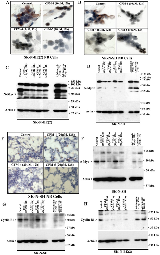
Figure 5. CFMs suppress expression of oncogenes N and c-myc.
(A, B, E) Cells were either untreated (Control), treated with indicated time and dose of respective CFMs, and followed by staining of cells using anti-N-myc (A, B) or c-myc (E) antibody as detailed in Methods. Presence of N-myc or c-myc proteins is indicated by intense brown staining in the nuclei of the untreated cells. (C, D, F-H) Cells were either untreated (Control) or treated with indicated agents for noted time and dose, and cell lysates were analyzed by Western blotting for levels of N-myc (C, D), c-myc (F), or cyclin B1 (panels G and H) and actin proteins as in Methods.

