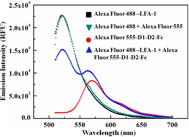
Figure 2. Ascertaining the FRET activity between Alexa Fluor 488-LFA-1 and Alexa Fluor 555-D1-D2-Fc.
Fluorescence emission spectra of individual fluorophore-protein conjugates, Alexa Fluor 488-LFA-1 (100 nM) and Alexa Fluor 555-D1-D2-Fc (100 nM), and a mixture of Alexa Fluor 488-LFA-1 (100 nM) and Alexa Fluor 555-D1-D2-Fc (100 nM) are compared. A sensitized acceptor emission at 570 nm and a donor quenching at 520 nm are observed in the emission spectra of the FRET mixture, confirming FRET. For control, the emission spectrum of the mixture of the dyes (Alexa Fluor 488+ Alexa Fluor 555), 100 nM each is also shown. That fact that no emission peak appeared at 570 nm for Alexa Fluor 555 when only the dye mixture (Alexa Fluor 488+ Alexa Fluor 488) was excited at 470 nm, whereas a prominent acceptor sensitized peak was observed for the FRET mixture (Alexa Fluor 488-LFA-1+ Alexa Fluor 555-D1-D2-Fc) indicates that the random FRET between the free dye molecules can be neglected in our study. All the spectra were obtained under the excitation of 470 nm. The gain of the spectrofluorometer was set at 100 (manual). The excitation and the emission bandwidths were fixed at 9 nm (for 316–850 nm excitation range) and 20 nm (for 280–850 nm emission range) for all the measurements. The fluorescence emissions were recorded with an integration time of 20 µs (more details on Table S1).

