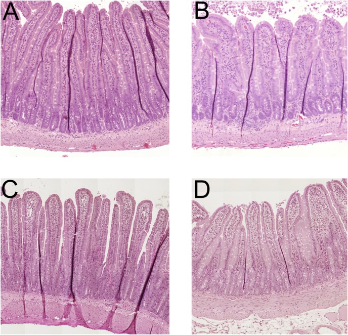
Figure 6. Histology of the duodenal mucosa.
The duodenum perfused with isotonic NaCl for 30(group I) had a normal morphological appearance (n = 4, Fig. 6A). The perfusion of the duodenal segment with 15% ethanol for 30 min (group II) caused mild villous tip damage observed as edema and the beginning of desquamation of the epithelium at the tip of less than 10% of the total villi (n = 4, Fig. 6B). The duodenal segment following perfusion of with 1.0 mM hydrochloric acid (pH 3) for 30 min (group III) had a normal morphological appearance (n = 4, Fig. 6C). The perfusion of the duodenal segment with 15% ethanol mixed in a hydrochloric acid solution of 1.0 mM for 30 min (group IV) caused mild villous tip damage observed as edema and the beginning of desquamation of the epithelium at the tip of less than 10% of the total villi (n = 4, Fig. 6D). The morphological changes in this group were not different from the group perfused with 15% ethanol alone.

