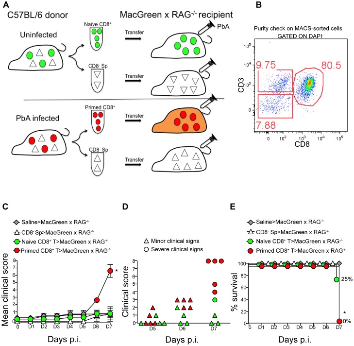
Figure 4. An adoptive transfer model to study the regulation of monocytes during ECM.
(A) CD8+ T cells or CD8− splenocyte fraction (Sp) isolated by MACS from uninfected or PbA-infected C57BL/6 donor mice on day 7 p.i. were adoptively transferred into PbA-infected MacGreen×RAG−/− recipient mice as depicted. Only primed CD8+ T cells (red circle) isolated from PbA-infected C57BL/6 donor mice induce NS (orange recipient). Representative data of 3 independent experiments is shown. (B) Dot plot shows purity of CD8+ T cells routinely obtained by MACS sorting. (C) MacGreen×RAG−/− recipient mice receiving saline (n = 4 mice), naïve (n = 4 mice) or primed CD8+ T cells (n = 5 mice) or CD8− splenocytes (n = 4 mice) were infected with PbA and clinical scores monitored daily, *p = 0.01 (unpaired t test). (D) Clinical score of each mouse on day 5–7 p.i. Each symbol represents one mouse. Recipients of naïve and primed CD8+ T cells are shown in green and red respectively. (E) Percent survival of MacGreen×RAG−/− recipients after adoptive transfer, *p<0.05 (Mantel-Cox test).

