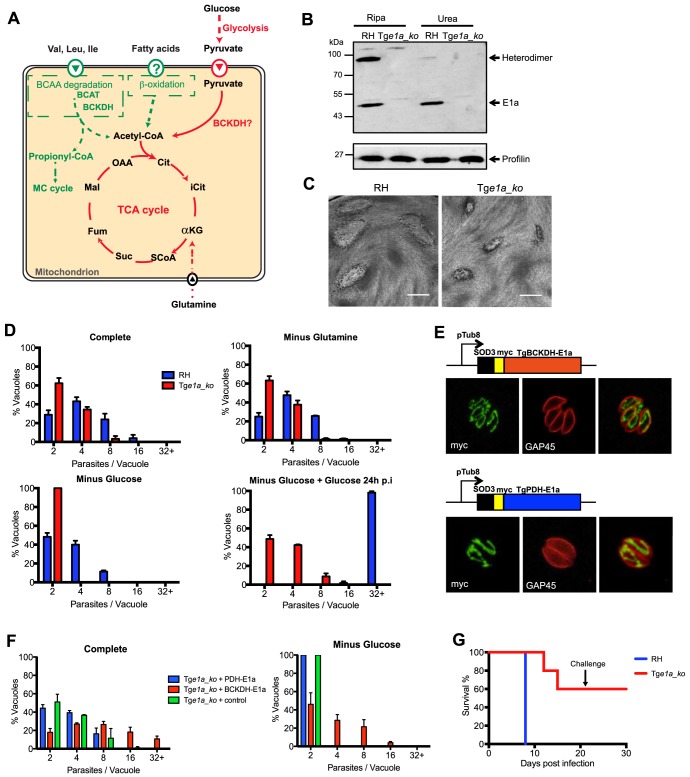
Figure 1. Toxoplasma gondii BCKDH-complex is required for normal growth and virulence.
(A) Schematic representation of pathways to produce acetyl-CoA in the mitochondrion. In green are pathways specific to T. gondii and in red pathways common to T. gondii and Plasmodium spp. (B) Total lysates from extracellular RHku80_ko (RH) and Tge1a_ko tachyzoites were analysed by Western blot. Expression of BCKDH-E1a was assessed using polyclonal anti-PfBCKDH-E1a antibodies. Detection of profilin was used as loading control. (C) Plaque assays were performed by inoculating HFF monolayers with RH or Tge1a_ko parasites for 7 days. Plaques were revealed by Giemsa staining of HFFs. Scale bar represents 1 mm. (D) Intracellular growth of RH (blue) and Tge1a_ko (red) was assessed after 24 h in complete media, media lacking glutamine, or glucose. Following 24 h of growth in glucose-depleted environment, glucose was added back to the media and rescue of the parasite’s growth was assessed. Data are represented as means ± SD from three independent biological replicates. (E) The apicoplast targeting sequence of TgPDH-E1a (aa 1–225, ABE76506) and mitochondrial targeting sequence of TgBCKDH-E1a (aa 1–73, XP_002366588) were replaced with the mitochondrial transit peptide of the superoxide dismutase 3 (SOD3) and myc-tagged [71] to direct the expression of the fusion protein in the mitochondrion of Tge1a_ko parasites for complementation (creating pTub8-SOD3mycPDHE1a and pTub8-SOD3mycBCKDHE1a respectively). Immunofluorescence assay shows localization of SOD3mycPDHE1a and SOD3mycBCKDHE1a in the single tubular mitochondrion (anti-myc (in green), anti-GAP45 (pellicle marker in red)). (F) Intracellular growth assay at 32 h post transient transfection of Tge1a_ko with pTub8-SOD3mycPDHE1a, pTub8-SOD3mycBCKDHE1a and pTub8-mycNtGAP45 (negative control) in complete media or media depleted in glucose. Data are represented as means ± SD from three independent biological replicates. Only vacuoles containing parasites transiently expressing the transgene were taken into account. Over 200 vacuoles were counted per replicate. (G) CD1 mice were infected with RH (in blue) or Tge1a_ko (in red) tachyzoites (∼15 parasites per mouse) and survival was assessed over 21 days. A challenge with ∼1000 wild-type RH tachyzoites was performed on mice that survived initial infection and survival followed for a further 10 days. Five mice were infected per condition.

