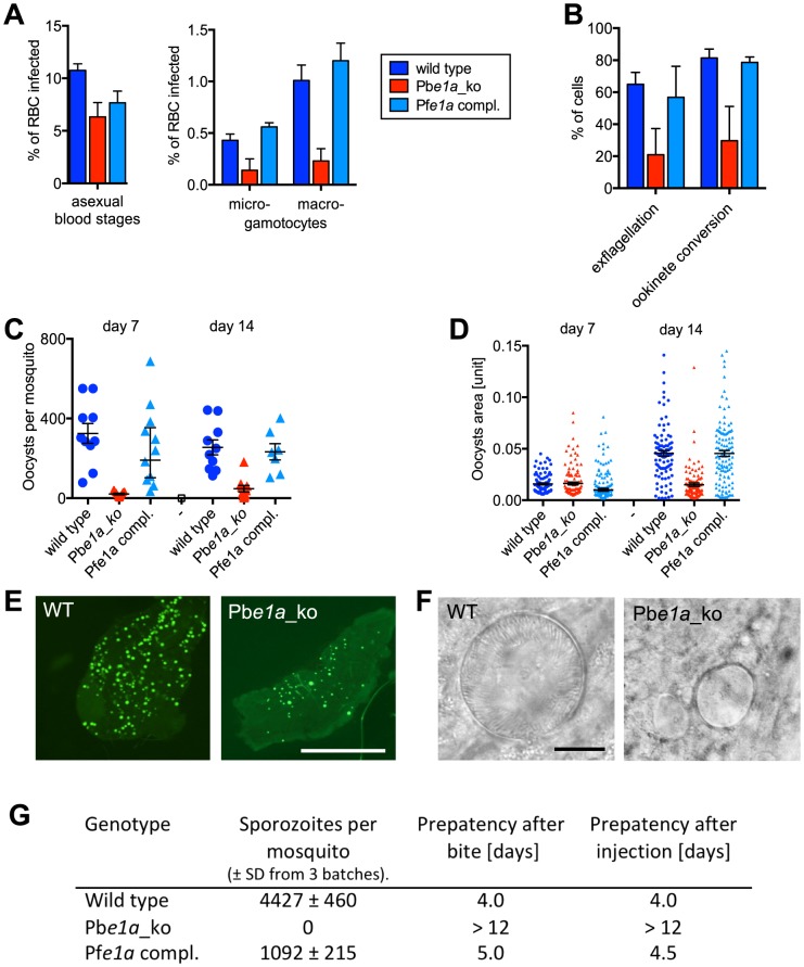
Figure 7. Role of BCKDH during sexual development and in mosquito stages of P. berghei.
(A) Asexual parasitaemia and male (micro-) and female (macro-) gametocytaemia in the peripheral blood of mice 3 days post infection with 1×10⁷ parasites i. p. Error bars show standard deviations from 3 mice. (B) Developmental capacity of gametocytes in vitro measured from the same infections shown in panel A. The relative ability of microgametocytes to release microgametes was assessed by counting exflagellation centres in a haemocytometer 15 min after addition to activating medium. The ability of activated macrogametocytes to become fertilised and convert to ookinetes was assessed by quantifying round and ookinete-shaped parasites following life staining of the surface marker P28. Colour code as in panel A; error bars show standard deviations. (C) Oocyst numbers on the midguts of individual A. stephensi mosquitoes on different days after feeding on three infected mice per mutant. Geometric means and 95% confidence intervals are also shown. (D) Sizes of individual oocysts from infected midguts at different days after infection. Black lines show geometric means and 95% confidence intervals. (E) Fluorescence micrographs of representative A. stephensi midguts dissected 14 days after feeding on wild type and mutant parasites expressing GFP. Scale bar = 0.5 mm. (F) Phase contrast images of representative oocysts. Scale bar = 10 µm. (G) Sporozoite numbers per mosquito as determined from 3 batches of 10 dissected salivary glands. Transmission from mosquito to mice was examined by measuring the prepatent period in 2 mice per group after bites from ∼20 mosquitoes or intraperitoneal injection of homogenates from 10 pairs of salivary glands. Data shown in all panels are representative of two independent experiments each performed with three infected mice per parasite line.

