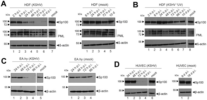
Figure 4. Soluble Sp100 levels are reduced upon KSHV infection of HDF, EA.hy and HUVEC cells.
Western blot analysis of low-salt soluble RIPA extracts prepared from: (A) HDF cells that had been infected with KSHV for the indicated time points (left panel, lanes 1–6) or mock infected cells at 0 h (left panel, lane 7) or between 8 h and 10 days post treatment (right panel, lanes 1–5), (B) mock infected HDF cells (lane 8) or HDF cultures exposed to UV-irradiated KSHV supernatants (lanes 1–7), (C) KSHV or mock infected EA.hy cells and (D) KSHV or mock infected HUVEC cells. β-actin served as a loading control in all panels.

