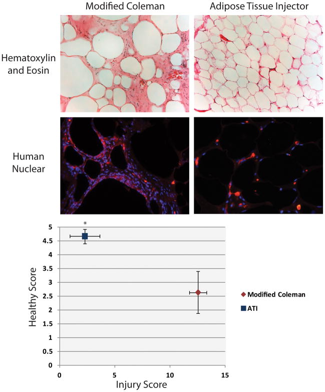
Figure 6.
Histological analysis of injected fat. A) Hematoxylin and eosin staining (top) of explanted fat 12 weeks after injection with modified Coleman technique (left) and ATI device (right). Human-Nuclear Antigen immunofluorescent staining (bottom) confirms human origin of explanted fat. Note increased infiltration of mouse-derived cells with modified Coleman technique. B) Chart of histological scoring with healthy score on y-axis and combined injury score (vacuoles, infiltrate, and fibrosis) on x-axis. Injected fat with ATI device (blue) had significantly greater healthy score and significantly lower injury score compared to modified Coleman technique (red) (*p < 0.05).

