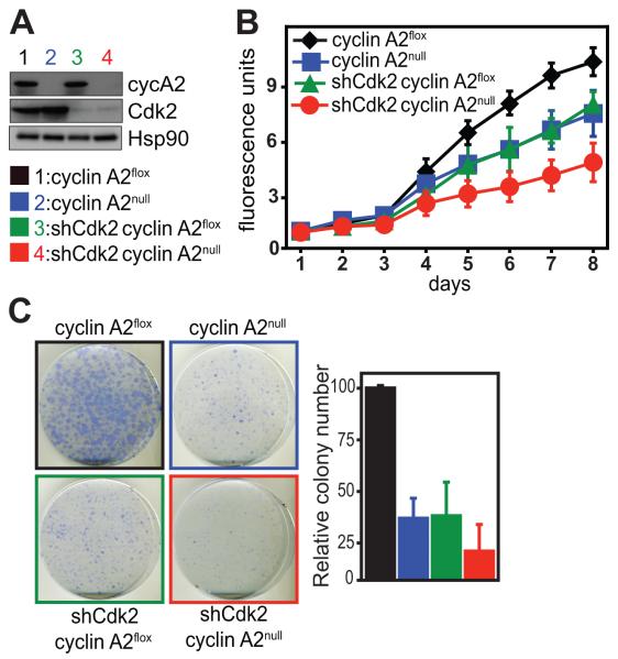
Fig. 3. Impaired proliferation of primary liver tumor cells upon inhibition of cyclin A2 and Cdk2.
Liver tumors were induced by tail vein injection of Ras/shRNA p53 in cyclin A2flox/floxRosa26-CreERT2Tg/Tg mice. Tumors were isolated followed by dissociation and culturing of tumor cells (A-C). Cyclin A2 knockout in primary liver tumor cells was achieved by addition of 4-OHT, whereas Cdk2 was silenced by retroviral shRNA transduction (A). Cyclin A2nullshCdk2 liver tumor cells exhibit decreased proliferation rates as determined by alamarBlue proliferation assay (B) and resistance to colony formation (C). Data in (A-C) is representative of two tumor cell lines established from two mice. NPIU: normalized phosphoimager units.

