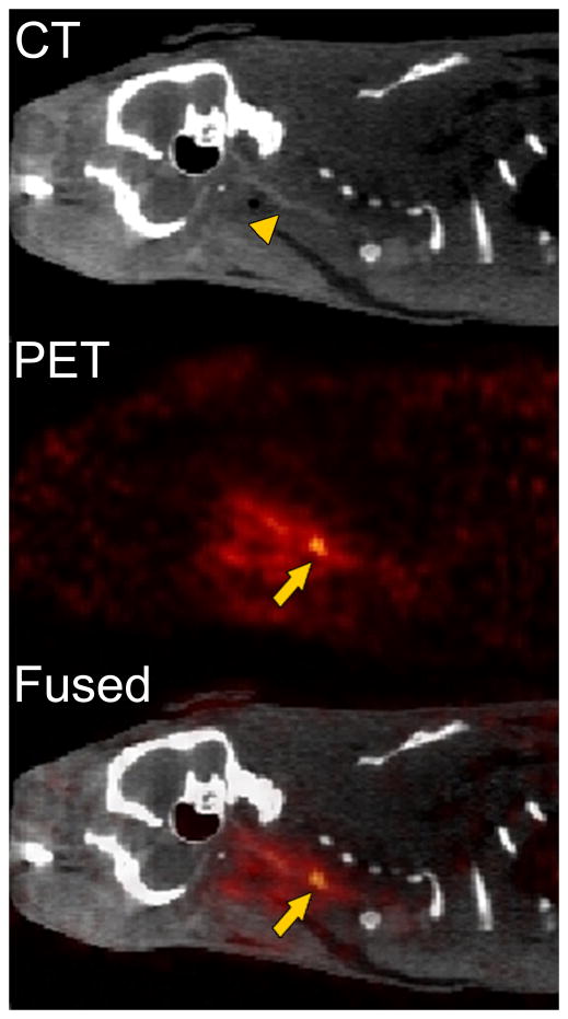
Figure 2.
Representative CT, PET, and fused PET-CT sagittal images reconstructed from data 240–285 min after the injection of FBP7 obtained from an animal that had carotid crush injury. FBP7 imaging reveals the thrombus localization at the level of the common carotid artery. Arrows indicate the crush injury site; arrowhead indicates the common carotid artery.

