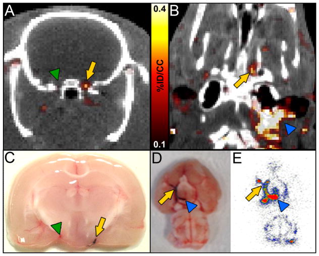
Figure 5.
PET-CT images showed that FBP7 detects occlusive thrombus in both intracranial (AB) and extracranial (B) arteries. A: arrow = ICA/MCA with thrombus; green arrowhead = contralateral ICA/MCA. B: arrow = intracranial ICA/MCA; blue arrowhead = extracranial ICA. Presence of thrombus was verified in postmortem brains from animals injected with Evans blue-labeled clot (C–D; arrow in C = ICA/MCA with thrombus; green arrowhead in C = contralateral ICA/MCA; arrow in D = intracranial ICA/MCA; blue arrowhead in D = extracranial ICA). Autoradiography replicated findings of PET-CT (E; arrow = intracranial ICA/MCA; blue arrowhead = extracranial ICA).

