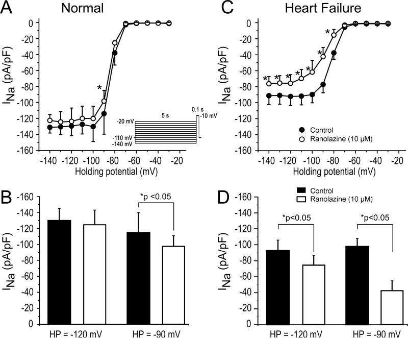
Figure 4.
Ranolazine causes a significantly greater reduction of peak cardiac sodium channel current (INa) in atrial myocytes isolated from heart failure (HF) vs. normal dogs. A: Relation showing magnitude of INa (at -10 mV) as a function of voltage for normal atrial cells (filled circles). Application of ranolazine (open circles) resulted in minor tonic block of INa. B: Bar graph highlighting the differences in INa magnitude at 2 different holding potentials (HPs) in the absence and presence of ranolazine. C: Relation showing size of INa (at -10 mV) as a function of voltage for HF atrial cells (filled circles). Application of ranolazine (open circles) resulted in significant tonic block of INa. D: Bar graph highlighting the differences in INa magnitude in HF atrial cells. The size of INa in HF atrial cells was reduced compared normal atrial cells and application of ranolazine resulted in greater tonic block. *p<0.05. n=6-8 (oneway repeated measures or multiple-comparison ANOVA followed by Bonferroni's test).

