
Table 2.
Echocardiographic measurements and myocardial values in patients with MD or DMD
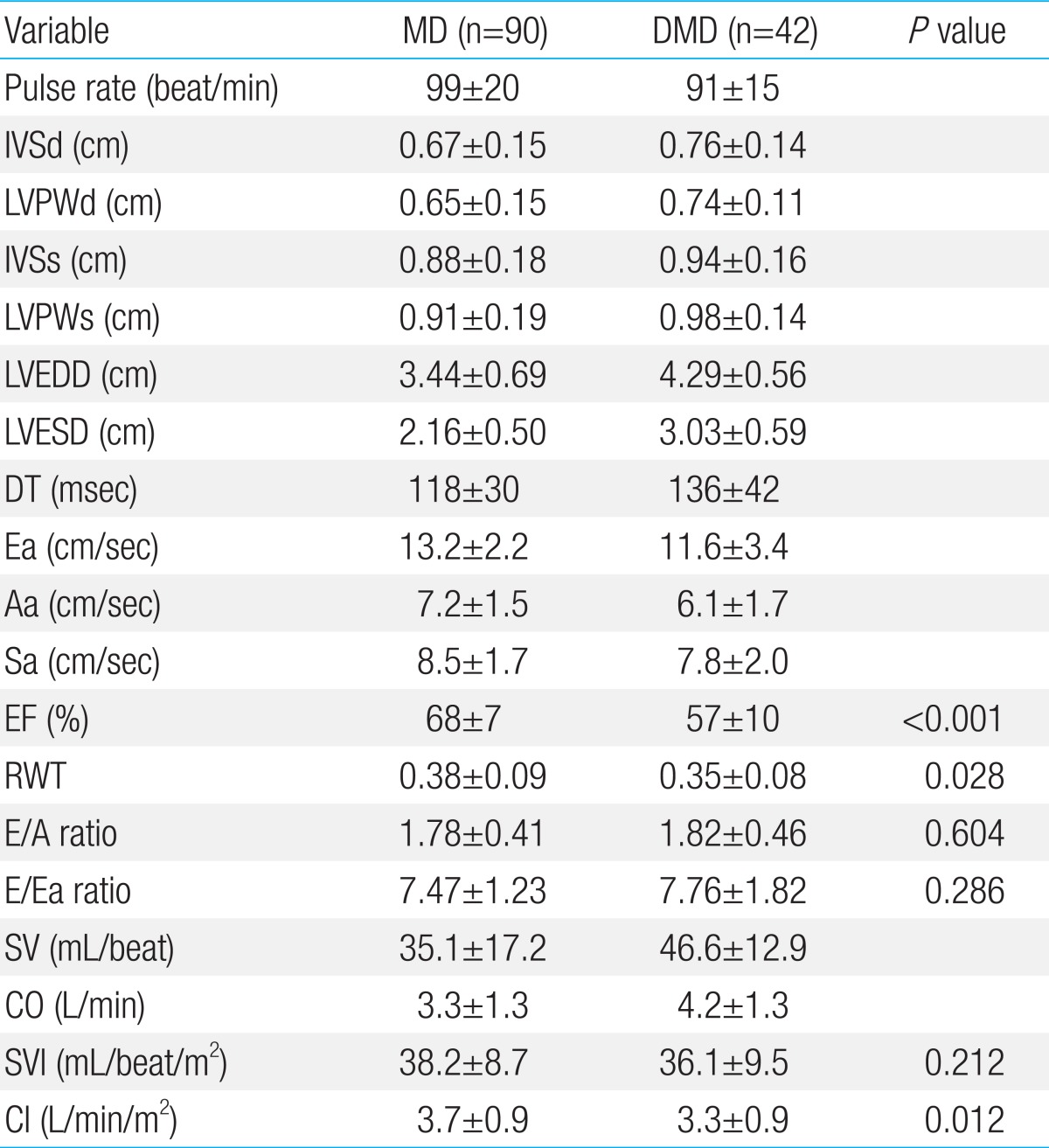
Values are presented as mean±standard deviation.
MD, mitochondrial disease; DMD, Duchenne muscular dystrophy; IVSd and LVPWd, interventricular septal thickness and left ventricular posterior wall thickness during the diastolic phase; IVSs and LVPWs, interventricular septal thickness and left ventricular posterior wall thickness during the systolic phase; LVEDD, left venticular end diastolic dimension; LVESD, left ventricular end systolic dimension; DT, deceleration time; Ea and Aa, early and late diastolic velocities of the mitral annulus; Sa, systolic peak velocity of the mitral annulus; EF, ejection fraction; RWT, relative wall thickness; E/A ratio, ratio of early to late mitral filling velocity; E/Ea ratio, ratio of early mitral filling velocity to early diastolic mitral annular velocity; SV, stroke volume; CO, cardiac output; SVI, stroke volume index; CI, cardiac index.