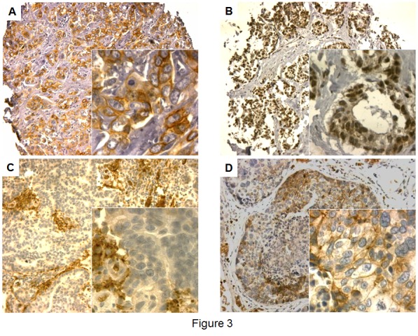
Figure 3. CADM1 immunostaining in primary breast cancer and BCBM samples.

A) Primary BC sample with homogenous positive CADM1 membrane and negative nuclear staining, B) Primary BC sample with negative CADM1 membrane and weak nuclear staining, C) BCBM sample with negative CADM1 membrane and nuclear staining with positively stained erythrocytes, D) BCBM sample with weak heterogeneous CADM1 membrane and negative nuclear staining.
