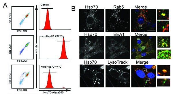
Fig 5. Exo-Hsp70 is internalized by cancer cells employing the mechanisms of active transport.

A. C6 cells incubated with Hsp70-Alexa Fluor®555 (50 µg/ml) for 18 h at various temperature regimens – 4°C or 37°C – were subjected to flow cytometry analysis. B. Confocal microscopy of C6 cells expressing marker proteins and incubated with Hsp70-Alexa®Fluor 488 (green) for 18 h. Upper panel: C6 cells tranfected with rab-5-RFP plasmid (red) after incubation with exo-Hsp70 (green); second line of images, C6 cells fixed, permeabilyzed and stained with anti-EEA-1 antibodies (red). Lower panel: lysomes were detected with the help of LysoTracker® (red), nuclei stained with DAPI (blue). Scale bar – 5 µm. Inserts show patterns of exo-Hsp70 staining co-localized and without vesicular structures.
