
Figure 5. JNJ-585 affects the repair response elicited by decitabine.
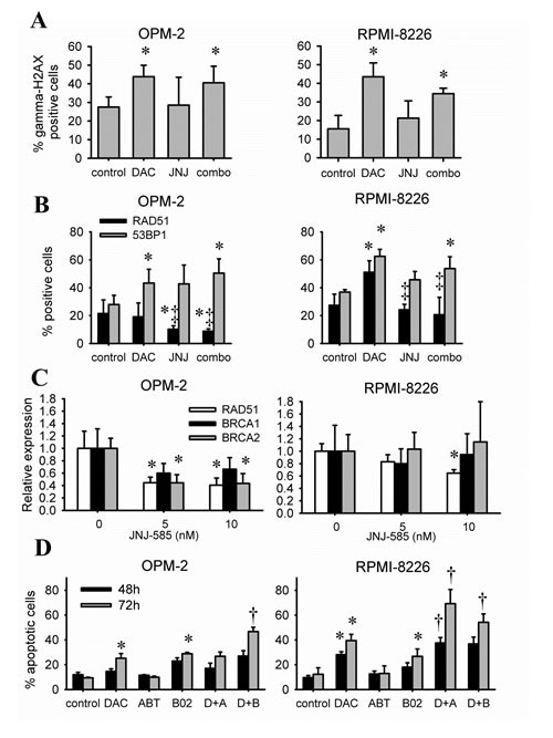
A-B: Cells were treated with decitabine (DAC) and/or JNJ-585 for 1 day. Doses for OPM-2 were 1µM decitabine and 2.5nM JNJ-585; for RPMI-8226 1µM decitabine and 5nM JNJ-585. Next, cytospins were made and stained for gamma-H2AX (A), RAD51 and 53BP1 (B). Images were quantified using ImageJ PZFociEZ plugin. A: Quantification of gamma-H2AX foci. B: Quantification of RAD51 and 53BP1 foci. At least 100 nuclei were analyzed and nuclei with at least 10 foci were scored as positive. C: Cells were treated with JNJ-585 for 1 day. Samples were processed and used for qRT-PCR to analyze expression of RAD51, BRCA1 and BRCA2. ABL-1 was used as housekeeping gene. D: Cells were treated with decitabine (1µM) and/or B02 (10µM) or ABT-888 (10µM) for 2 or 3 days. Next, apoptosis was determined by flow cytometry using AnnexinV-FITC/7'AAD staining. Apoptotic cell percentage is the sum of annexinV+ and AnnexinV+/7'AAD+ cell percentage. Data is shown as mean ± SD of 3 experiments. * indicates p<0.05 compared to untreated conditions. ‡ indicates p<0.05 compared to decitabine. † indicates p<0.05 compared to single agents.