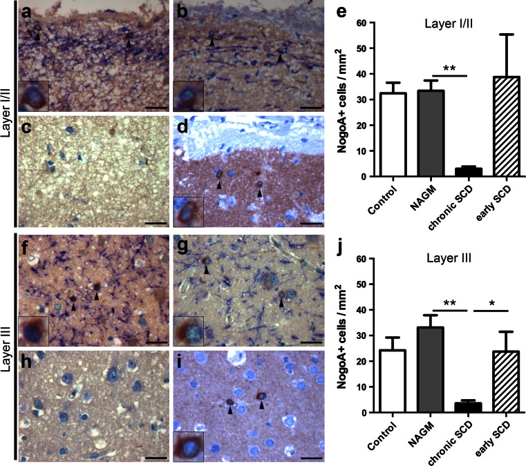
Fig. 3.
Reduction of oligodendrocytes in cortical layers I/II and layer III in chronic, but not early MS lesions. a–j Immunohistochemistry for MBP (blue) and NogoA+ OLs (brown) within layers I/II (a–d) or layer III (f–i), respectively, and the corresponding quantitative evaluation (e, j). Representative images of control cortex (a, f), normal-appearing cortex (NAGM) of chronic MS (b, g) as well as chronic (c, h) or early (d, i) SCD. Similar numbers of NogoA+ cells are noted in cortical layers I/II (a, b, d, e) and layer III (f, g, i, j) in the control cortex (a, f), NAGM (b, g) and SCD from patients with early MS (d, i). In patients with chronic MS, NogoA+ cells are significantly reduced in chronic SCD (c, h) in cortical layers I/II (c) and III (h) each compared to the corresponding NAGM area. The densities of NogoA+ OLs in layer III are significantly decreased in chronic SCD compared to early SCD (j), whereas there is a trend for the group comparison within layers I/II (e) (p = 0.06). NogoA+ cells are indicated by black arrowheads and shown in detail in insets. Scale bars 25 μm in a–d and f–i; error bars indicate SEM. *p < 0.05, **p < 0.01

