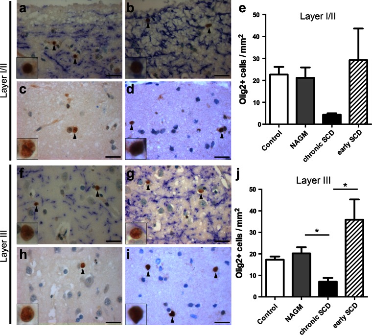
Fig. 4.
Reduced oligodendrocyte precursors in chronic, but not early MS lesions. a–j Immunohistochemistry for OPCs with strong Olig2 expression (Olig2+ cells) (brown) and MBP-positive myelin (blue) within layers I/II (a–d) or layer III (f–i), respectively, and the corresponding quantitative evaluation (e, j). Representative images are shown of control cortex (a, f), normal-appearing cortex (NAGM) of chronic MS (b, g) as well as chronic (c, h) and early (d, i) SCD. Similar numbers of Olig2+ cells are noted in myelinated cortical layers I/II (e) or layer III (j) in control cortex, MS NAGM and SCD from patients with early MS. In layers I/II, there is a trend for decreased Olig2+ OPCs in chronic SCD (autopsy) (c) compared to either adjacent NAGM (p = 0.05) or demyelinated early SCD (biopsy) (p = 0.06) (e). In layer III Olig2+ cells are significantly reduced in chronic SCD (h, j) compared to NAGM and early SCD (p < 0.05 for both comparisons). Olig2+ cells are indicated by black arrowheads and shown in detail in insets. Scale bars 25 μm in a–d and f–i; error bars indicate SEM. *p < 0.05

