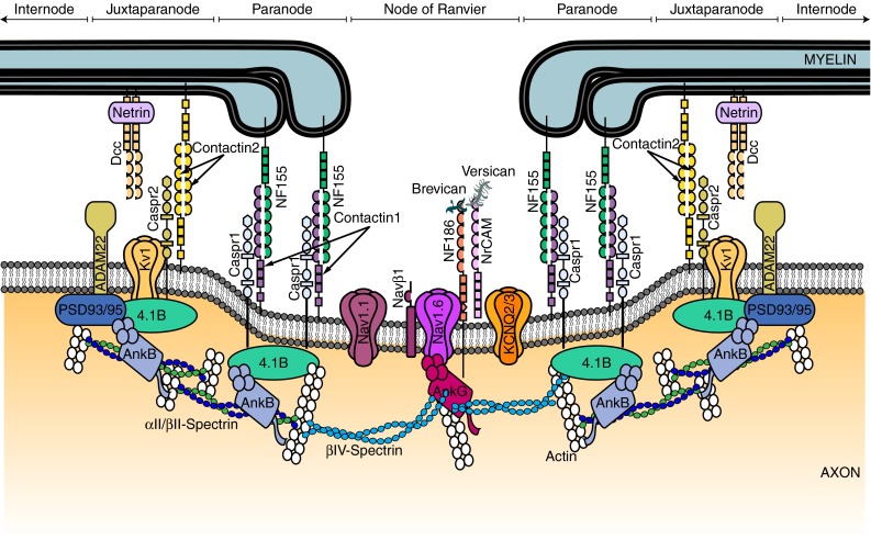
Fig. 2.

Schematic diagram of the proteins at the node of Ranvier, paranode and juxtaparanode. These domains are the location of ion channels (Nav1.6 and Nav1.1, KCNQ2/3, Kv3.1 and Kv1.1/1.2), cell adhesion molecules (neurofascin 155 (NF155), neurofascin 186 (NF186), contactin 1 and 2, contactin-associated protein (Caspr 1 and 2), cytoskeletal scaffolding proteins [Ankyrin (Ank) G and B, protein 4.1B, and postsynaptic density protein 93/95 (PSD93/95)], cytoskeletal proteins (βII- and βIV-spectrin), and extracellular matrix proteins (brevican, versican and a secreted form of NrCAM). Targeting and scaffolding mechanisms ensure that each protein is segregated to its specific subdomain
