
Fig. 4. PDK inhibition by phenylbutyrate.
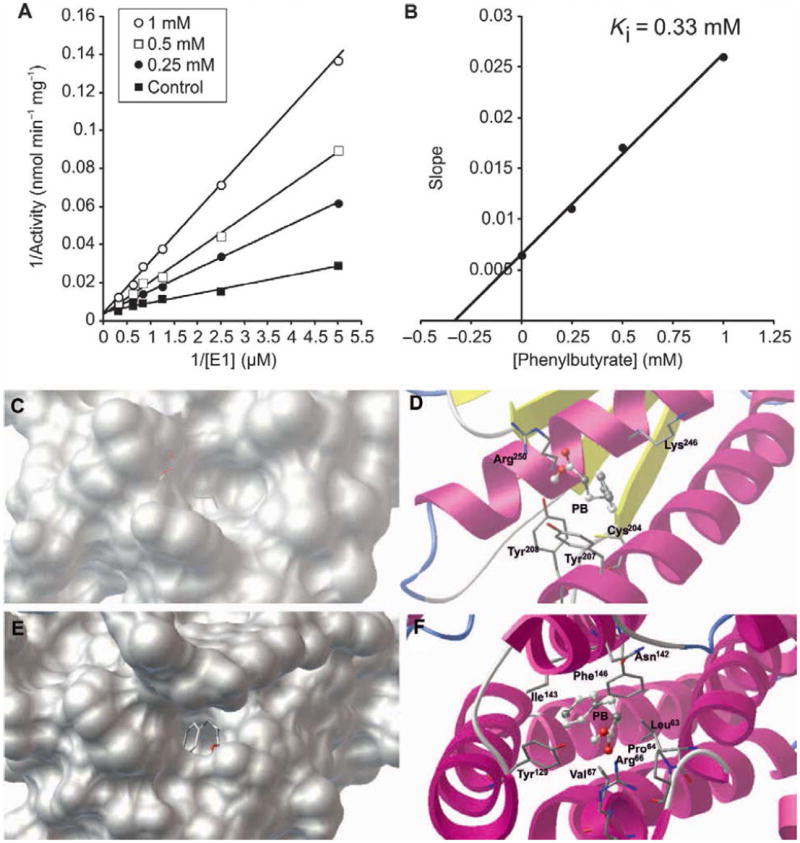
A. Lineweaver-Burk plot of WT recombinant E1α in the absence (■) or in the presence of 0.25 mM (●), 0.5 mM (□), and 1 mM (○) of phenylbutyrate. B. Replot of slopes measured from (A) against the concentration of phenylbutyrate. The intersection of the line with the x axis gives the value of Ki. C and E. Phenylbutyrate (PB, stick representation) in the two identified pockets on the protein surface of PDK2. D and F. Specific interactions of phenylbutyrate with amino acid residues (stick representation) at the binding sites. Van der Waals interaction spheres of the amino acid residues (stick representation) in contact with the inhibitor have been removed for clarity.