Figure 4.
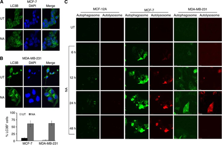
Nimocinol acetate induces autophagosome and autolysosome formation in BCa cells. (A and B) Confocal microscopy images showing autophagosome formation, marked by the presence of LC3B puncta. (C) MCF-12A, MCF-7, and MDA-MB-231 cells were treated with NA and images of the cells were captured using a live cell imaging microscope programmed to take photographs every 1 h, for up to 48 h. Autophagosomes, with neutral pH, are indicated by acid-sensitive GFP emission, whereas autolysosomes, with acidic pH, are indicated by acid-insensitive RFP emission concomitant with loss of GFP emission.

