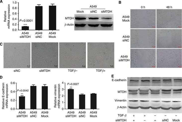
Figure 5.
Knockdown of MTDH expression increases migration in A549 cells. (A) Q-PCR and western blotting analyses for expression of MTDH in selected knockdown A549 cells (A549 siMTDH) by transfecting MTDH-targeting siRNA. Negative control siRNA-transfected A549 cells (A549 siNC) and only lipo-transfected A549 cells (A549 Mock) served as negative control. GAPDH and β-actin were, respectively, used as control for mRNA and protein. ***P<0.0001 compared with control. (B) Migration ability of A549 cells. Migration ability of A549 siMTDH was significantly decreased than that in A549 siNC and A549 Mock. (C) Cell morphology of A549 cells. Typical polygonal epithelial structure in A549 siNC was transformed into slender, reduced cell adhesive, spindle-shaped mesenchymal-like structure in A549 siMTDH cells, which was similar to the cell morphology after TGF-β stimulation. (D) Q-PCR analysis of EMT markers' mRNA expression in A549 cells. The mRNA expression of E-cadherin was significantly decreased and that of Vimentin increased in A549 siMTDH cells. Data are mean±s.d. *P<0.05, **P<0.01 compared with control. (E) Western blotting analysis of EMT markers' protein expression in A549 cells. E-cadherin expression was decreased and Vimentin expression was increased in A549 siMTDH cells and was also similar to the changes after TGF-β stimulation. The downtrend of E-cadherin expression and uptrend of Vimentin expression was increased after TGF-β stimulation in A549 siMTDH cells.

