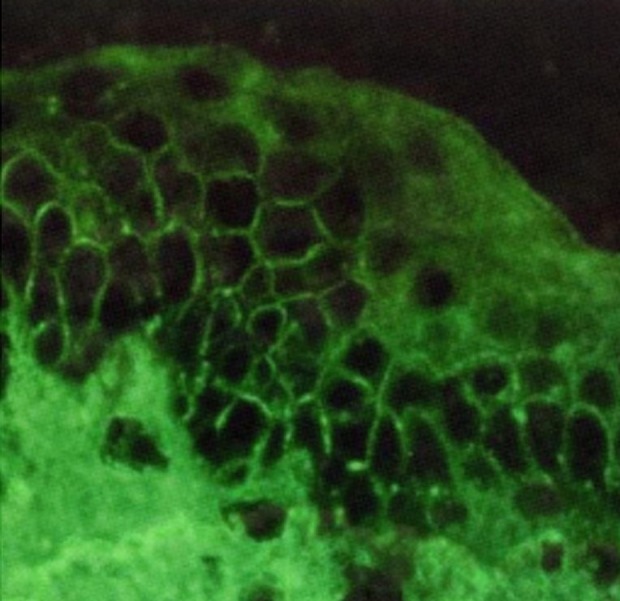
Figure 4. Direct immunofluorescence microscopy (DIF) of the cutaneous lesions revealed weak in vivo IgG deposition on the keratinocyte cell surface from the mid to upper epidermal layers (weakly positive lace like pattern in the epidermis, original magnification x100).

