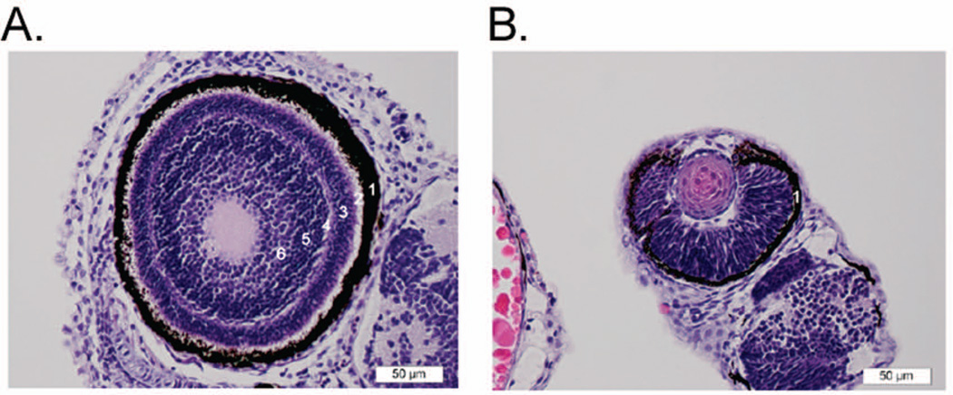
Figure 7.
Marcks is involved in retinal histogenesis of zebrafish. (A) Normal eye from 72 hpf wild type zebrafish embryo. Here, the layers of the retina are relatively well defined. Starting from the outermost layer and working toward the center of the eye: 1) the dark brown/black retinal pigment epithelium; 2) layer of rods and cones; 3) outer nuclear layer (dark blue/purple); 4) outer plexiform layer; 5) inner nuclear layer; 6) inner plexiform layer. The ganglion cell and nerve fiber layers are less distinct in this section. The plexiform layers consist mainly of synapses of the various sensory neural cells. (B) Eye from 72 hpf MAT injected zebrafish embryo (moderate phenotype). The retina consists of a mass of poorly organized neural cell nuclei with no clear separation into normal retinal layers. Only the retinal pigment epithelium forms a distinct layer (1). This morphology is consistent in both the MAT and MBT injected fish, and is most severe in the severe phenotype fish (data not shown).

