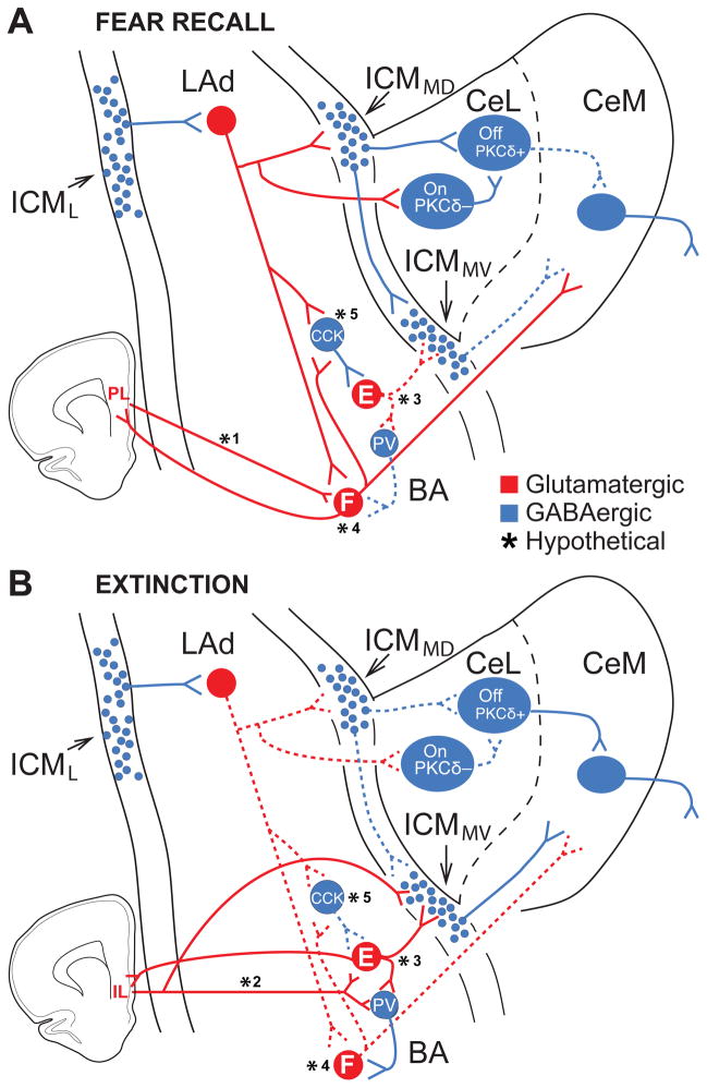
Fig. 3.
Intra-amygdala interactions supporting expression and extinction of conditioned fear. The model includes known pathways and hypothetical links (marked by asterisks) that collectively account for most of the available evidence. Solid and dashed lines represent connections that are more or less active, respectively. (A) The increased CS responsiveness of CeM output neurons after conditioning likely results from two parallel mechanisms: excitation by glutamatergic BA neurons plus disinhibition from CeL and ICMMV inputs. CeM excitation: CS-induced LA activation causes a BA neuron subtype (“Fear neurons”, F) to fire and excite CeM cells whereas another type of BA neurons (“Extinction cells”, E) are inhibited, possibly by CCK+ interneurons. Although LA neurons respond transiently to the CS, BA fear neurons, through excitatory interactions with each other and/or with prelimbic (PL) cells (lower left), would prolong the transient tone signal emanating from LA into persistent CS responses. CeM disinhibition: The excitation of LA cells also leads to the recruitment of ICMMD neurons and of a subgroup of CeL cells, likely PKCδ− (CeL-On) cells. ICMMD neurons would then inhibit ICMMV cells, disinhibiting CeM neurons. In addition, ICMMD cells would inhibit subgroups of CeL neurons, possibly PKCδ+ (CeL-Off) cells. The recruitment of PKCδ− (CeL-On) cells by LAd neurons would cause a further inhibition of PKCδ+-neurons and disinhibition of CeM cells. (B) The decreased CS responsiveness of CeM output neurons after extinction likely depends on two parallel mechanisms: disfacilitation of CeM cells and increased feedforward inhibition of CeM neurons. CeM disfacilitation: The rapid extinction of LAd responses to the CS results in a diminished recruitment of BA fear neurons, disfacilitation of CeM neurons, and, possibly, of CCK+ interneurons. As a result, BA extinction cells are disinhibited. Reciprocal excitatory interactions between IL (lower left) and BA might also contribute to enhance the excitability of extinction cells. The disinhibition of extinction cells causes increased excitation of a different set of BA interneurons, possibly PV+ interneurons, controlling fear cells. CeM inhibition: the reduced CS responsiveness of LAd neurons would cause a disfacilitation of ICMMD neurons and consequent disinhibition of ICMMV neurons. This effect would coincide with an increased excitation of ICMMV cells by inputs from BA extinction cells, thus resulting in an increased feedforward inhibition of CeM cells. The disfacilitation of ICMMD neurons would also cause a disinhibition of subsets of CeL cells, possibly corresponding to PKCδ+ neurons. This effect would be reinforced by the reduced activation of PKCδ− cells secondary to reduced LAd inputs. Hypothetical connections (marked by asterisks). (*1, *2) While it was shown that BA fear and extinction cells differentially project to PL and IL, respectively, whether return mPFC projections are similarly segregated is unknown. (*3, *4) Currently, there is no data available on the connections of fear and extinction neurons with other amygdala neurons. The differential connections shown are hypotheses based on the available literature. (*5) Little data is available on the connectivity of CCK cells with different types of BA neurons. Trouche et al. (2013) reported that they contact fear (not shown here) and extinction-resistant neurons. CCK synapses to extinction-resistant (but not fear neurons) showed an up-regulation of CB1 receptors after extinction training. The input and output connections of CCK interneurons shown in the figure are all hypothetical. It is possible that other subtypes of interneurons are differentially connected to fear and extinction neurons.

