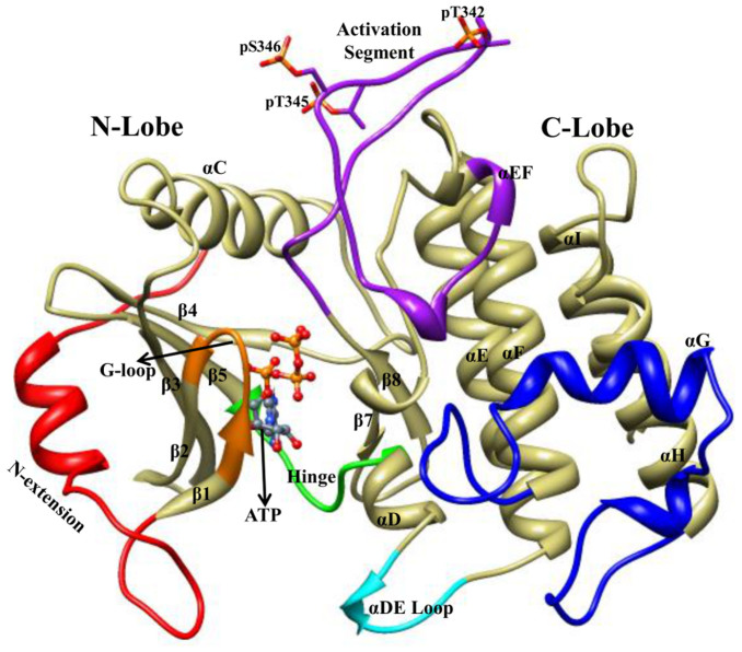
Figure 1. Structure of IRAK4 kinase domain.
The structure of IRAK4 KD predominantly shows the N lobe with β-strands and an α-helix, C lobe with exclusively α-helices. The structural parts were shown in various colors, N-terminal extension preceding kinase domain in red, G loop in orange, activation segment in magenta, hinge region in green, αDE loop in cyan, helix αG and its adjoining loops in blue. ATP molecule between the N and C lobe were shown in ball and stick model in slate gray color. Phosphorylated residues pT342, pT345, and pS346 are highlighted. The structural parts were labeled according to the crystal structure32.

