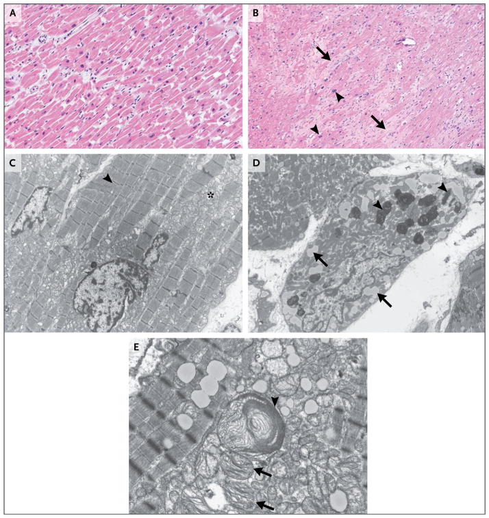
Figure 2. Micrographs of Specimens of Apical Left Ventricular Tissue.
Panel A shows a photomicrograph of a specimen of normal myocardium (hematoxylin and eosin) obtained from an autopsy specimen. Panel B shows a photomicrograph of a specimen of the patient’s left ventricular apical core (hematoxylin and eosin) obtained during implantation of a left ventricular assist device for cardiogenic shock. Moderate interstitial, pericellular fibrosis (arrows) and myocyte hypertrophy (arrowheads) are evident. Panel C shows a transmission electron micrograph of an autopsy specimen. Normal myocytes with abundant myofibrils (arrowhead) and morphologically normal mitochondria (asterisk) are present. Panel D shows a transmission electron micrograph of the patient’s left ventricular tissue. Myocytes have degenerative features, characterized by substantial loss of contractile units, intracytoplasmic lipid accumulation (arrows), and lipofuscin deposition (arrowheads). Panel E shows another transmission electron micrograph of the patient’s left ventricular tissue. Both a highly atypical, enlarged mitochondrion (arrowhead) and immediately beneath it several smaller mitochondria (arrows) containing abnormally configured cristae suggest direct mitochondrial injury.

