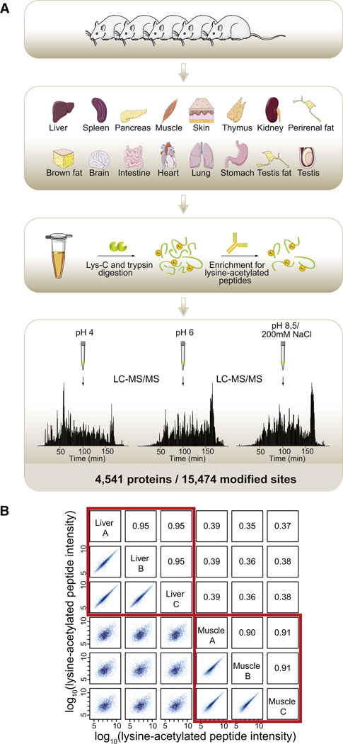
Figure 1. Workflow for Acetylome Analysis of Rat Tissues.
(A) A total of 16 tissues were isolated from 5 male rats; the tissues were snap frozen, heat inactivated, homogenized, and solubilized. The extracted proteins were digested with endoproteinase Lys-C and trypsin, and lysine-acetylated peptides were enriched by immunoprecipitation. The acetylated peptide mixtures were fractionated by SCX in a STAGE tip, and three pH elutions per tissue were analyzed by high-resolution LC-MS/MS on a LTQ-Orbitrap Velos instrument resulting in identification of a total of 15,474 lysine acetylation sites from 4,541 proteins.
(B) For liver and muscle samples, results from three technical replicates prepared from the tissue homogenates are shown. Logarithmized intensities for acetylated peptides were plotted against each other and shown on the left side of the diagonal with the corresponding Pearson correlation coefficients given on the right side of the diagonal. Technical replicates of the same tissue are highly correlated.
See also Figures S1 and S2.

