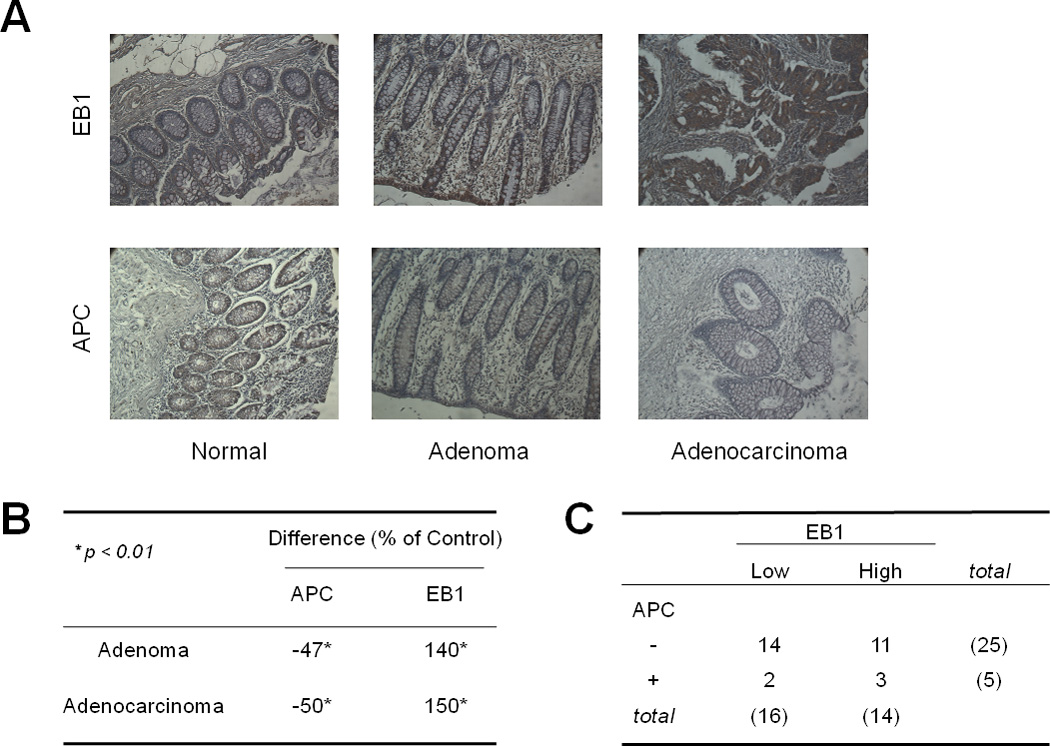
Figure 1. Immunohistochemical staining of EB1 and APC during human colon cancer progression.

A) Expression of EB1 and APC (c-terminus) in human normal, adenoma, and adenocarcinoma colonic tissues. B) Quantification of the expression of EB1 and APC in adenoma and adenocarcinoma samples compared to normal tissue, *p <0.01. C) Correlation between EB1 and APC in adenoma tissue. High expression indicates an intensity score of 3 or higher.
