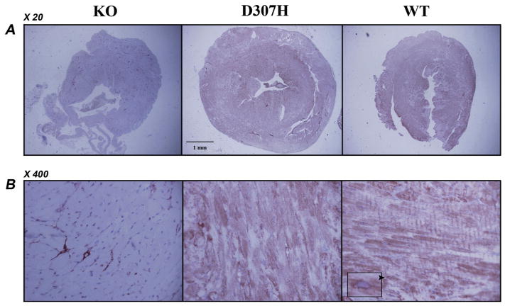
Fig. 3.
Immunohistochemistry in hearts of CASQ2 mutant mice. Representative sections of mouse hearts stained with anti-calsequestrin antibody X20 (A) and X400 (B). Diffuse staining for CASQ2 and striations are prominent in WT heart. A predominantly perinuclear distribution may be noted (arrowhead and insert). KO hearts has no staining apart from the capillary network, which is attributable to cross reactivity with CASQ1 in the smooth muscle. D307H had a non-homogenous positive staining in the myocardium. D307H, CASQ2D307H/D307H; KO, CASQ2Δ/Δ; WT, wild type; CASQ, calsequestrin. n = 3 hearts/group, 2 sections/heart, animal age 7–8 month.

