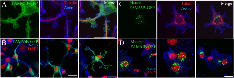
Fig. 4.
Effect of the FAM65B Δ34–86 mutation on subcellular localization. (A and B) In transfected COS7 cells, the green fluorescence corresponding to wild-type FAM65B-GFP defines the plasma membrane of the cell as the cortical actin network does (in blue) and is also present in the intracellular membrane compartment (A, higher magnification; B, overview of cell groups). (C and D) Localization of mutant FAM65B-GFP to cytoplasmic inclusion bodies and not to membranes (D, overview of cell groups). The microtubule network of the cell cytoskeleton is visualized using an antibody against β-tubulin (A and D), whereas the cellular nuclei are stained with DAPI. (Scale bars, 10 µm.)

