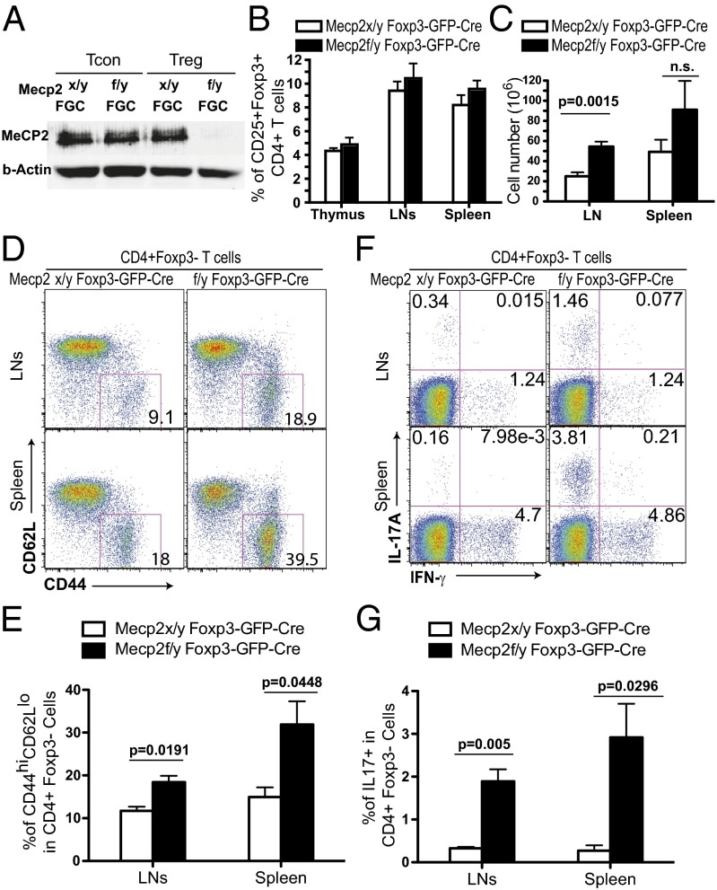
Fig. 1.
Mild immune activation in young adult Mecp2f/y Foxp3–GFP–Cre mice. (A) MeCP2 expression was examined by Western blot in sorted CD4+CD25−GFP− Tcon and CD4+CD25+GFP+ Treg cells from the spleens and LNs of Mecp2x/y Foxp3–GFP–Cre or Mecp2f/y Foxp3–GFP–Cre mice. (B and C) Lymphocytes from the thymus, LNs, and spleens of 8–10-wk-old Mecp2f/y Foxp3–GFP–Cre or littermate control mice were isolated for enumeration of cell numbers and flow cytometry analysis. Data are shown as means ± SEM (n = 5). (B) Percentage of CD25+Foxp3+ cells in CD4+ T cells. (C) Absolute cell numbers in the LNs and spleens. (D and E) Expression of CD44 and CD62L on the surface of CD4+Foxp3− T cells from LNs and spleens. (F and G) Lymphocytes isolated freshly ex vivo were stained for intracellular cytokines after 4 h of stimulation with 0.9 nM PdBU and 0.5 μg/mL ionomycin in the presence of 5 μg/mL brefeldin A and 2 μM monensin.

