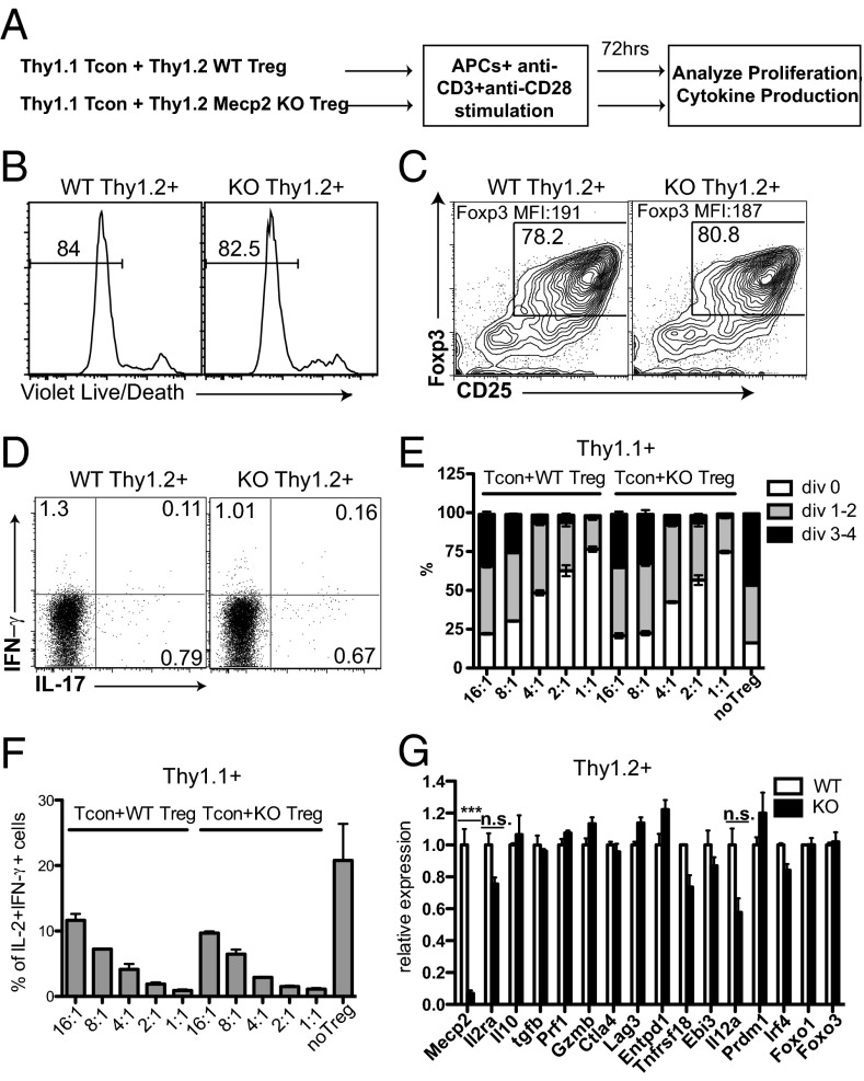
Fig. 3.
MeCP2-deficient Tregs are competent in suppressing effector T-cell activation during short-term culture in vitro. CD4+CD25− conventional T cells from LNs and spleens of Thy1.1+ B6 mice were FACS sorted, labeled with CFSE, mixed with Thy1.2+ CD4+CD25+ Tregs from LNs and spleens of Mecp2f/y Lck–Cre or littermate control mice at the indicated ratios, and stimulated with 0.5 μg/mL anti-CD3 and 0.5 μg/mL anti-CD28 in the presence of T-cell–depleted splenocytes (as APCs) from B6 mice for 72 h. (A) Schematic representation of the workflow. (B–D) The survival (B), Foxp3 maintenance (C), and cytokine production (D) from WT and MeCP2-deficient Tregs (Thy1.2+) as shown by LIVE/DEAD Fixable Violet Dead Cell Staining and intracellular staining, respectively. Data shown represent three independent experiments. (E and F) The suppression of effector T-cell (Thy1.1+) proliferation (E) and cytokine production (F) by MeCP2-sufficient or -deficient Tregs (Thy1.2+) as shown by CFSE dilution and intracellular staining. Bar graphs show means ± SEM of three independent experiments. (G) Viable WT and MeCP2-deficient Thy1.2+ Treg cells from the 4:1 Tcon:Treg culture were sorted, and expression of genes that are critical for Treg suppressive function was determined by qPCR. Data show means ± SEM of two independent experiments.

