Fig. 6.
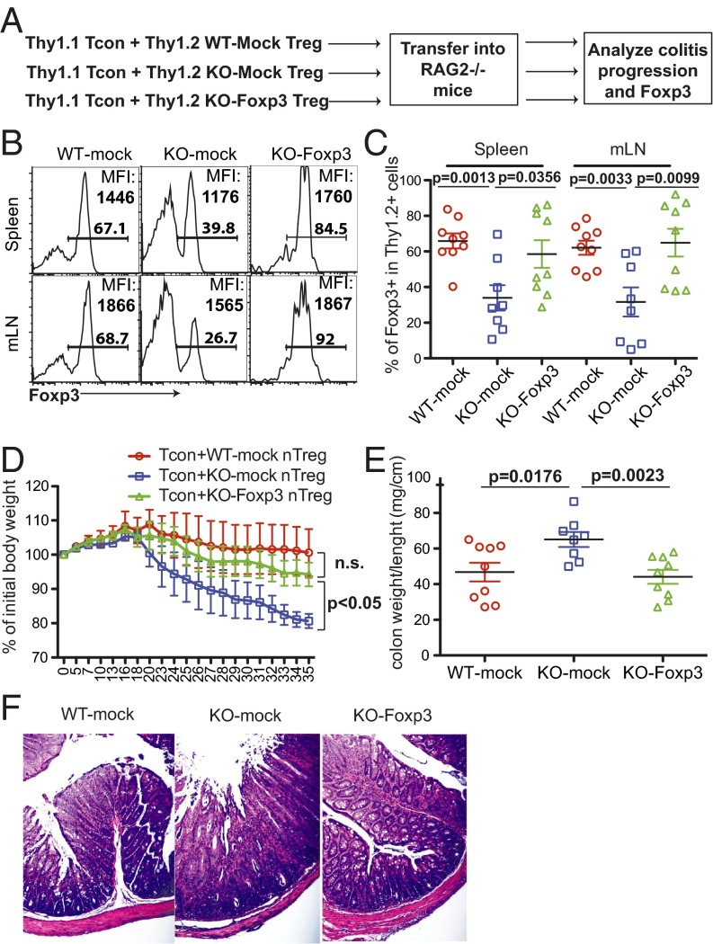
Ectopic expression of Foxp3 restores the capacity of MeCP2-deficient Tregs to suppress effector T-cell–mediated colitis in vivo. Thy1.2+ CD4+CD25+ Tregs from LNs and spleens of Mecp2f/y Lck–Cre or their WT littermate control mice were sorted and stimulated with 1 μg/mL anti-CD3 and anti-CD28 in the presence of 50 U/mL IL-2. Eighteen hours later, cells were transduced with a retrovirus that encodes the foxp3 coding sequence together with GFP (Foxp3) or GFP alone (mock). Two days later, 5.0 × 10⁴ Thy1.2+GFP+ Tregs were sorted and mixed with 5.0 × 10⁵ naïve conventional T cells (CD25− CD45RBhi CD4+) from WT Thy1.1+ B6 donors, and then transferred into RAG2−/− mice. Colon pathology and phenotype of transferred cells were analyzed 5 wk after adoptive transfer (n ≥ 8). (A) Schematic view of the workflow. (B and C) Percentage of recovered Foxp3+ cells from the Thy1.2+ origin and MFI of Foxp3 expression in Foxp3+ Tregs. (B) Representative FACS plots. (C) Summary of results from various experimental groups. Each symbol represents one single recipient mouse. (D) Weight changes during the colitis progression. The weight of recipients at different time points was normalized to the initial body weight of the individual mouse before transfer. Data show means ± SEM. (E and F) Colon immunopathology of recipient mice was indicated by the ratio of colon weight to length (E) and illustrated by H&E staining (F).

