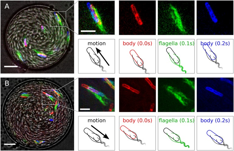
Fig. 3.
Drop overview in gray scale: bright field image. Positions of the membrane (false colored red at time t = 0, blue at t = 0.2 s) and flagella (false colored green at t = 0.1 s) dyes help determine the cell orientation. (A) Forward motion: cell at the oil interface both point and move to the top left corner. (B) Backward motion: the cell is pointing to the top left corner while moving overall in the opposite direction. (Scale bars: at the drop images, 10 μm; at the individual bacterium images, 5 μm.)

