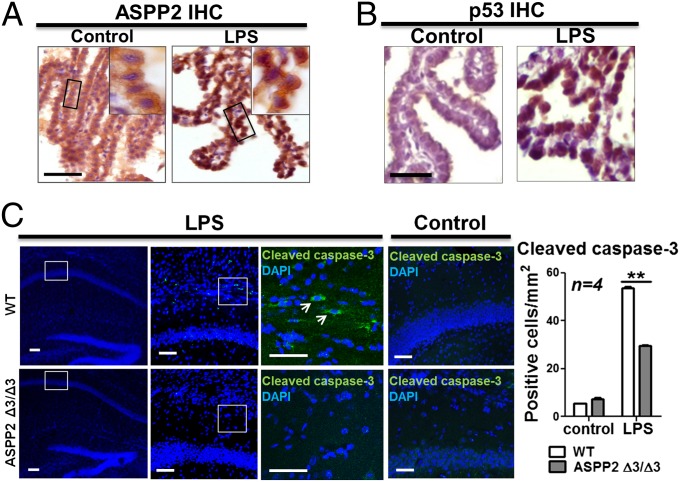
Fig. 3.
LPS induces nuclear ASPP2 expression in a model of maternal inflammation, and ASPP2 mediates apoptosis. (A) On LPS injection, ASPP2 is disrupted from the TJs and relocalized to the nucleus of CP epithelial cells. (Scale bar: 25 µm.) (B) After LPS injection, p53 appears in the nucleus of CP epithelial cells. (Scale bar: 25 µm.) (C) IF staining of cleaved caspse-3 in LPS-injected and control saline-injected WT and ASPP2 Δ3/Δ3 mice. Arrows indicate cleaved caspase-3–positive cells. (Scale bars: 25 µm.) Quantification of the number of cleaved caspase-3–positive cells in the hippocampus after LPS or saline injection (n = 4). **P < 0.01.

