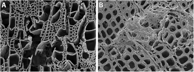
Fig. 3.

Wood decay experiments indicating mode of decay by Botryobasidium botryosum and Jaapia argillacea. (A) Micrograph of B. botryosum on aspen wood with vessel, fiber, and parenchyma cell walls degraded. Mycelia are visible growing through the voids. (B) Micrograph of J. argillacea on pine showing an area within the wood where the fungus has caused a localized simultaneous decay of the cells. Residual cell wall material and mycelia fill the degraded zone.
