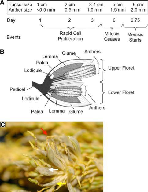
Fig. 3.
Landmark development stages in initial tassel and anther ontogeny. a Approximately 29 days are required for anther initiation within spikelets to pollen shed (Ma et al. 2008). Over the course of approximately 1 week, tassels grow from 1 to 6 cm in length. At the earliest stage, anther primordia are present, and these expand to 0.5 mm by the next day. Within anthers, mitotic proliferation continues until day 6, the 1.5-mm stage, when all cell types are present in a normal anther. After 18 h, meiosis starts in the central cells (Ma et al. 2008). b Progression of tumor location within a spikelet. Spikelets are encased by a pair of leaf-like glumes and contain two florets. Each floret contains three stamens (subtending filament plus anther), encased by a palea and a lemma. The lodicules are generally considered to be modified petals (Bommert et al. 2005); their expansion at spikelet maturity separates the glumes, permitting emergence of mature anthers. Infections of tassels <1 cm results in conversion of the entire spikelet to a tumor, with few recognizable maize structures. Infections at the 1-cm tassel stage result in tumors primarily in the palea and lemma plus stamen filaments, and from the 2–4-cm-tassel stage, upper floret filaments and anthers are converted to tumorous growths. Infections after cessation of cell division in anthers (day 6 in a) can cause tumors in the lodicules. c Tumors and abnormal stamens in the apical region of a W23 tassel induced by injection at the ~1-cm tassel stage. Entire spikelets are converted to tumors (middle arrow), and in some cases recognizable stamens emerge from the tip of such converted spikelets (upper arrow). In some florets, U. maydis infection in the filament is readily visualized (lower arrow), and the anther has an abnormal morphology

