
Fig. 1.
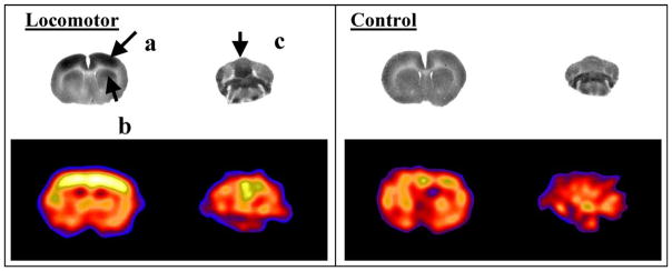
Changes in cerebral blood flow-related tissue radioactivity elicited during treadmill walking in the rat compared to a quiescent control. In the locomotor paradigm, the tracer was injected while the rat was walking for 5 min on a RotaRod treadmill. Injection during the control condition occurred while the animal was resting quietly in a transport cage placed next to the RotaRod. Row 1: Coronal autoradiographs of the distribution of [14C]-iodoantipyrine tissue radioactivity show activation of (a) primary motor cortex, (b) dorsolateral striatum, as well as (c) midline cerebellum in the exercising animal [108]. Row 2: Coronal section in a rat obtained using a micro-PET R4 Rodent scanner (Concorde Microsystems, Inc., Knoxville, TN) after administration of [64Cu]-PTSM (1.25 mCi/kg), show a similar activation pattern to that obtained with [14C]-iodoantipyrine. Increased CBF tissue radioactivity is depicted in warmer colors. Brightness was normalized between the two rat PET studies based on a defined region within the piriform cortex, which did not display any significant change in response to the treadmill paradigm. Enhanced resolution of the PET images was obtained using maximum-a-posteriori (MAP) reconstruction [136,137].