
Figure 6. Changes in FGF signaling cannot induce cell fate switch after PrE and EPI specification.
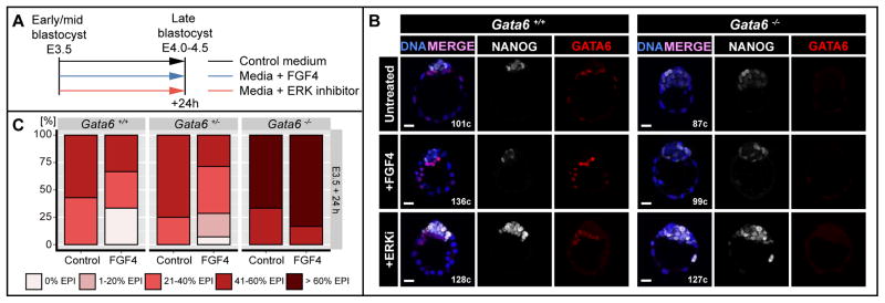
A Experimental timeline. B Representative immunofluorescence images of Gata6+/+ and Gata6−/− embryos cultured from the salt-and-pepper (E3.5) stage for 24h in medium without (untreated) or with 500ng/ml FGF4 + 1μg/ml Heparin (+FGF4) or medium + 1μM PD0325901 (ERKi). Blue: Hoechst; white: NANOG; red: GATA6. C Percentages of embryos falling into 1 of five FGF-response categories: 0% EPI, 1–20% EPI, 21–40% EPI, 41–60% EPI and >60% EPI in ICMs of embryos treated as in (A). EPI: Epiblast.