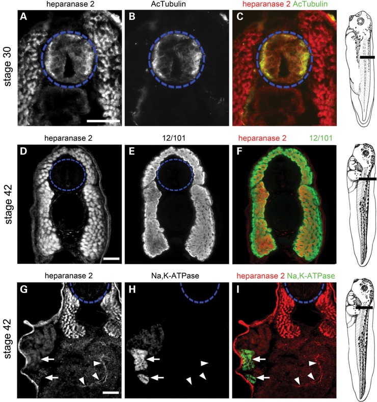
Figure 3.
Tissue-specific localization of heparanase 2. Transverse sections through the trunk of Xenopus embryos, as indicated in the diagrams shown on the right, imaged by immunofluorescence, with the neural tube outlined by blue dashed circles. In merged images, heparanase 2 localization is indicated in red while other proteins are indicated in green. (A–C) Heparanase 2 colocalized with acetylated α-tubulin (AcTubulin) in the lateral zones of the Stage 30 neural tube. The flanking myotomes were also positive for heparanase 2. (D–F) At Stage 42, heparanase 2 was absent in the neural tube but myotomes, co-immunostained with the muscle-marker antibody 12/101 (it is the name of an antibody), remained positive. (G–I) The Stage 42 pronephric tubule did not display a specific IHC signal for heparanase 2; note that here, the weak signal is background autofluorescence. The two arrows indicate a proximal tubule which in H and I is seen to be reactive with Na+/K+-ATPase antibody. The set of three arrowheads in (G)–(I) demonstrate specific heparanase 2 immunostaining in the apical zone of epithelia lining the gut lumen. Scale bars are 50 µm.

