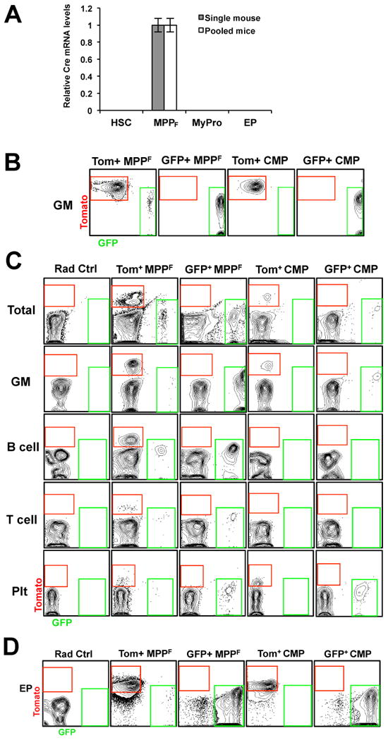
Figure 2. Lack of Cre expression and activity in myeloid progenitors at steady-state and upon in vivo and in vitro differentiation.

(A) Quantitative RT-PCR analyses of Cre recombinase mRNA levels in Flk2+ (MPPF) and Flk2- (HSC; myeloid progenitors (MyPro; Lin-c-kit+Sca1- cells), and erythroid progenitors (EP; Ter119-Mac1-Gr1-B220-CD3-CD71+)) cell populations from FlkSwitch mice revealed that only MPPF express Cre. Bar graph indicates the relative levels of Cre mRNA in cell populations sorted from individual (grey bar) or multiple (white bar; n=3) FlkSwitch mice. β-actin was used as a positive control for all populations. Error bars indicate SEM. (B-D) Tom+ CMP remain unfloxed during myeloid development. (B) Flow cytometry analysis of CMP progeny after 10 days of in vitro methylcellulose culture (n=6 in 2 independent experiments). (C) Tom and GFP analysis of donor-derived nucleated cells (total), GM, B-cells, T-cells, and Plts in PB of sublethally irradiated mice transplanted with Tomato+ or GFP+ MPPF (800 per mouse) or CMP (10,000 per mouse) (n=5-7 in 2 independent experiments). (D) Erythroid progenitor (EP) readout in spleens of lethally irradiated mice 9 days post-transplantation of Tom+ or GFP+ MPPF or CMP (n=10 in 2 independent experiments).
