Figure 2.
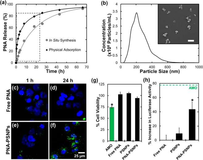
(a) Cumulative release of physically adsorbed PNA and in situ synthesized PNA in PBS at 37 °C. Release of in situ synthesized PNA was significantly lower (p < 0.005) for time points within the boxed region. (b) PNA–PSNP size distribution characterized by NTA. Inset: Representative SEM image demonstrating PSNP size and morphology (scale bar = 400 nm). (c–f) Confocal micrographs of Huh7 cells incubated for either 1 or 24 h with (c and d) free PNA, or (e and f) PNA–PSNPs at a 2 μM dose of anti-miR-122 PNA (3.7 μg/mL PSNPs for PNA–PSNP treatments). Labels: Hoechst nuclear dye (blue), Alexa-488 labeled PNA (green). (g) PSNPs had no significant effect on Huh7 cell viability relative to untreated negative control samples, in contrast to a 2′OMe-modified AMO delivered with Fugene 6 (p < 0.05). (h) Anti-miR122 activity indicated by luciferase activity in Huh7 psiCHECK-miR122 cells 44 h after treatment with free PNA, empty PSNPs, or PNA–PSNPs (2 μM dose of anti-miR-122 PNA, 3.7 μg/mL PSNPs). Results are normalized to cell number and expressed relative to luciferase activity in Huh7 psiCHECK-miR122 control cells without treatment. Dashed green line indicates anti-miR122 activity 44 h after treatment with 2 μM of an optimized anti-miR-122 AMO34 delivered via the Fugene 6 commercial transfection reagent.

