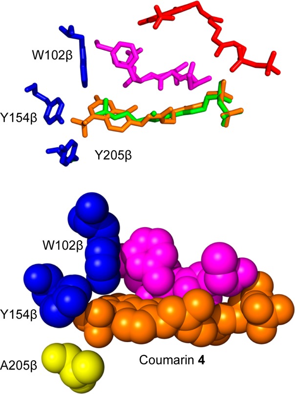
Figure 7.

Computational modeling structures of coumarin analogues in PFTase. (Above) Overlays of coumarin analogue docked in WT structure (red), courmarin analogue docked in Y205A mutant (orange), and FPP bound in the WT structure (green). The peptide substrate (purple) and the three relevant amino acids (blue) are also displayed with the surface of the WT enzyme. (Below) Coumarin analogue docked into Y205A mutant binding pocket. The structure is shown as spheres to show the coumarin moiety binding into the place occupied by the tyrosine residue in the WT structure. Coumarin analogue showed in orange, peptides substrate shown in purple, residues W102β and Y154β shown in blue, and the mutated Y205β shown in yellow.
