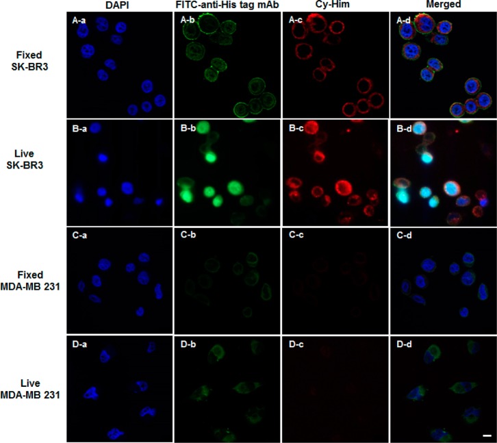
Figure 4.
Confocal fluorescence microscope images of HER2 labeling in HER2-positive SK-BR3 and HER2-negative MDA-MB-231 cells. Fixed cells were incubated with scFv-L-Aff (7 μg/mL) for 1 h at room temperature, followed by consecutive incubations with FITC–anti-His tag mAb and Cy-Him (1 μM) for 30 min each at room temperature. Live cells were treated with scFv-L-Aff and Cy-Him consecutively. After fixation, scFv-L-Aff was stained with FITC-anti-His tag mAb. The nucleus was counterstained with 4′,6-diamidino-2-phenylindole (DAPI) (scale bar = 10 μm).

