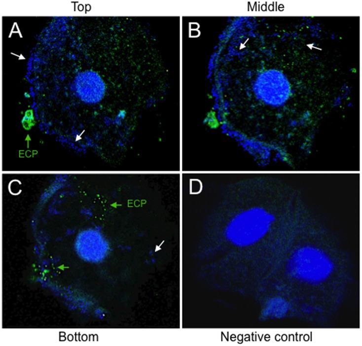
Figure 7. Confocal microscopy of an exfoliated bladder epithelial cell obtained from the midstream urine of a patient with UTI.
ECP production was detected by confocal microscopy in urine samples of women clinically diagnosed with acute UTI. (A, B, C) The presence of ECP (green arrows, Alexa-Fluor 488) on the bacteria (white arrows, bacteria stained blue with DAPI) present on exfoliated epithelial cells was demonstrated using an anti-EcpA antibody (1:1,500). Note the presence of extra- and intracellular bacteria in the different layers (A, top; B, middle; C, bottom) obtained by confocal microscopy. (D) An urine sample from a clinically healthy woman was used as a negative control. Images were taken with a 40× objective of Zeiss LMS-510 laser scanning microscope.

