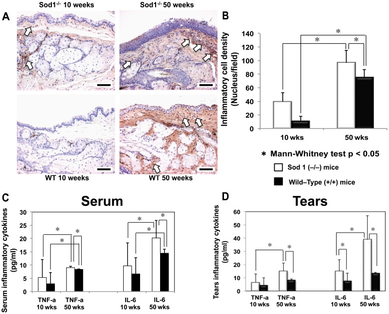
Figure 3. Inflammatory changes in the meibomian glands, serum and tears of the Sod1 −/− and wild type mice.
A, Specimens stained with CD45 from 10 and 50 weeks Cu, Zn-Superoxide Dismutase-1 knockout (Sod1 −/−) and wild type (WT) mice. Note the timewise increase in inflammatory cell staining from 10 to 50 weeks in the Sod1 −/− mice. Wild type mice specimens showed scant inflammatory cell staining, which increased significantly in the 50 week WT mice but not to the extent observed in the Sod1 −/− mice at 50 weeks. Bar = 50 micrometer. B, A significant timewise increase in the mean inflammatory cell densities from 10 to 50 weeks was observed in both the Sod1 −/− and WT mice (p = 0.0143 and p = 0.0286, respectively). Note the significantly higher inflammatory cell density in the Sod1 −/− mice at 50 weeks compared to the age matched WT mice (p = 0.0317). Five tissue sections of each eye of 6 animals (5 per mouse eye) were analyzed to produce the figure. Data represent the mean ± standard deviation for 6 mice from the Sod1 −/− groups and 6 mice from the WT groups at 10 and 50 weeks. C, The mean serum IL-6 concentration increased significantly from 10 to 50 weeks in both the Sod1 −/− (p = 0.0195) and WT mice (p = 0.0001). Serum IL-6 concentration was significantly higher in the 50 week Sod1 −/− compared to 50 week WT mice (p = 0.0100). Serum TNF-α levels were also significantly higher (p = 0.0011) in the 50 week Sod1 −/− mice compared to the age matched WT mice. The mean serum TNF-α concentration significantly increased from 10 to 50 weeks in the Sod1 −/− mice (p = 0.0457). Data represent the mean ± standard deviation for 14 and 8 Sod1 −/− mice at 10 and 50 weeks as well as 11 and 9 wild type mice at 10 and 50 weeks, respectively. D, There was also a significant (p = 0.0148) timewise increase in the mean tear IL-6 concentration in the Sod1 −/− mice from 10 to 50 weeks. Note the significantly higher IL-6 concentration in the Sod1 −/− mice at 10 (p = 0.0414) and 50 weeks (p = 0.0022) compared to the age matched WT mice. Tear TNF-α concentrations increased significantly from 10 to 50 weeks in the Sod1 −/− (p = 0.0115). Also note the significantly higher TNF-α concentration in the Sod1 −/− mice compared to the WT mice at 50 weeks (p = 0.0087). Data represent the mean ± standard deviation for 7 mice from the Sod1 −/− groups and 9 mice from the WT groups at 10 and 50 weeks.

