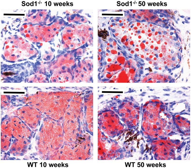
Figure 5. Timewise alterations in meibomian gland lipid staining.
Oil Red O staining showed an accumulation of large lipid droplets in the Cu, Zn-Superoxide Dismutase-1 knockout (Sod1 −/−) mice from 10 to 50 weeks. Note the diffusely uniform staining pattern in the wild type (WT) mice at 10 weeks and some accumulation of large lipid droplets in the WT mice at 50 weeks, but not to the extent observed in the same age Sod1 −/− mice. Bar = 100 micrometer. Five tissues sections from each eye of the 7 animals in each mice group were analyzed to produce the representative images.

